Fluoroscopy
With the aid of a contrast agent, Fluoroscopy enables a x-ray technologist to capture an image of an internal body organ while it is functioning. This contrast agent allows the image to be viewed clearly on a monitor or screen.
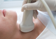
Ultrasound
Ultrasound imaging, also called sonography, is a method of obtaining diagnostic images from inside the human body through the use of high frequency sound waves. Utrasonography is used as a diagnostic tool that can assist doctors with making recommendations for further treatment.
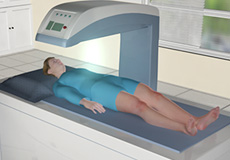
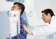
DEXA (Bone Densitometry)
To accurately detect osteoporosis, doctors commonly use DEXA bone densitometry to measure bone mineral density (BMD). DEXA is a quick, painless procedure for measuring bone loss. Measurement of the lower spine and hips are most often done.