Hampton Roads’ Premier Radiology
70 Years of Trusted Precision and Expertise in Radiology
Delivering expert, minimally invasive care—24/7, 365 days a year
70 Years of Trusted Precision and Expertise in Radiology
Delivering expert, minimally invasive care—24/7, 365 days a year
Expert Radiology Care Led by Physicians
Patient-centered imaging guided by experience and clinical excellence
Our practice is independently physician owned, allowing our radiologists to focus fully on patient care and diagnostic accuracy. Every study is interpreted by board certified physicians who work closely with referring providers to ensure clear communication and dependable results. With advanced imaging technology and a commitment to quality and safety, we deliver radiology services patients can trust at every stage of care.
interventional radiology
Minimally Invasive, Image-Guided Care for Faster, Safer Recovery
At Medical Center Radiologists, our expert, board certified interventional radiologists serve Hampton Roads with minimally invasive treatments—ranging from cancer care and pain relief to neurointerventional procedures—all delivered with advanced imaging technology, fewer complications and often no hospital stay.
Cancer treatments for liver, lung, kidney & bone
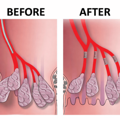
Men’s and women’s advanced health solutions including PAE & UAE treatment
Neurointerventional excellence: stroke, aneurysm, AVMs with pioneering techniques since 1996

Excellence in Interventional Radiology at Medical Center Radiologists
Medical Center Radiologists is nationally recognized for leadership in interventional radiology, including advanced ablations, TACE, and Y90 procedures. Based on AcuityMD data from 2020–2025, MCR is ranked #1 in the Mid-Atlantic region, #5 on the East Coast, and #13 across the United States. Our board certified interventional radiologists use the latest minimally invasive techniques to treat complex conditions with precision, compassion and excellent clinical outcomes.
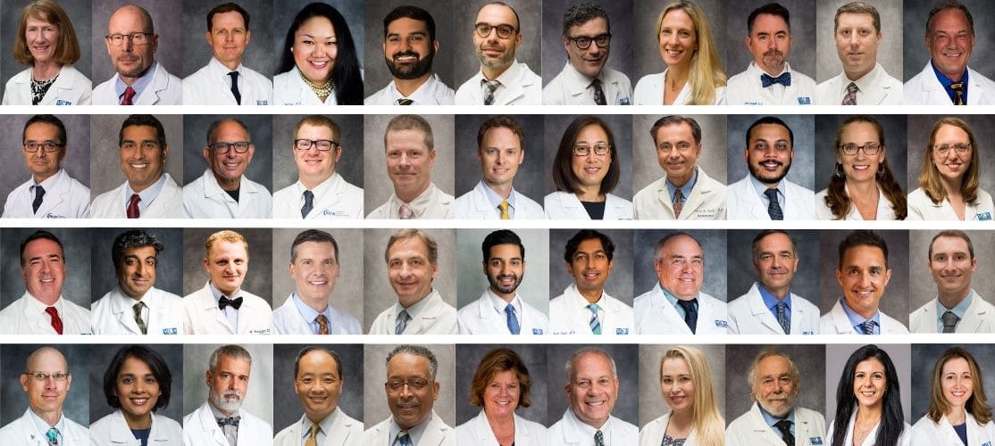
Meet Our Team
Tidewater’s Trusted Radiologists
Medical Center Radiologists (MCR) is the largest radiology practice in Southeast Virginia, proudly serving Hampton Roads for over 70 years. Our team of fellowship-trained physicians offers expertise across multiple subspecialties, ensuring precise diagnoses and advanced treatment options. As faculty at Virginia Medical School at Old Dominion University, our doctors are committed to educating future radiologists while delivering compassionate, patient-focused care to our community.
patient testimonials
5-Star Reviews From Real Patients
“Cannot say enough about the results of the PAE performed by the doctors at the Medical Center Radiologists. My quality of life has put me back at what it was at least 30 years ago. Saw results in about a week and very little uncomfortable pain the 1st 24 hours. Would recommend any one with prostate problems to look into this procedure.”
“This procedure after 30 days took me off all medication related to the prostate. I feel like I did in my early 20s. Went from 5x a night to 1x every so often. It’s like a miracle procedure. I normally don’t do surveys but this is something I wanted to recommend.” -Robert
“The professionals at Sentara General Hospital’s Interventional Radiology department were incredibly helpful and kind. I received thorough explanations during each visit, and my questions were always answered with patience and respect. I am so happy I found them.”


our locations